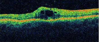
Optical Coherence Tomography (OCT) is a noninvasive imaging technique that has been used increasingly to diagnose and manage a variety of retinal diseases and glaucoma. OCT is based on the principal of Michelson interferometry. Interference patterns produced by low coherence light reflected from retinal tissues and a reference mirror are processed into an “A-scan” signal. Multiple A-scan signals are aligned to produce a two-dimensional image that can be thought of as a form of “in vivo histology.” Optical coherence tomography has been used to identify macular holes, to differentiate macular holes from simulating lesions, to identify lamellar macular holes, macular cysts, vitreomacular traction, subretinal fluid, pigment epithelial detachment, and choroidal neovascularization. It can be used to identify and quantify macular edema, and to measure retinal thickness changes in response to therapy. Macular thickness measurements determined by OCT correlate well with visual acuity and with leakage observed by fluorescein angiography. Optical coherence tomography is an accurate and reproducible method to measure retinal nerve fiber layer thickness. it can be used to differentiate glaucomatous eyes from normal eyes.
Optical Coherence Tomography